Hypertrophic Pulmonary Osteoarthropathy
General Considerations
- Clinical syndrome consisting of clubbing of the fingers and toes
- Painful and swollen joints
- Symmetric periostitis especially involving the tibia and fibula, radius and ulna
- Most often secondary to an intrathoracic neoplasm or infection
- Primary form is called pachydermoperiostitis
Etiology
- Release of vasodilators which are not metabolized by lung
- Increased flow through AV shunts
- Reflex peripheral vasodilation (vagal impulses)
- Hormones: estrogen, growth hormone, prostaglandin
Thoracic Causes
- Malignant tumor (0.7-12%)
- Benign tumor
- Chronic infection / inflammation
- Congenital heart disease with R-to-L shunt
Extrathoracic Causes
- GI tract
- Liver disease
- Biliary + alcoholic cirrhosis
- Posthepatic cirrhosis
- Chronic active hepatitis
- Bile duct carcinoma
- Benign bile duct stricture
- Amyloidosis, liver
- Abscess
- Undifferentiated nasopharyngeal carcinoma
- Pancreatic carcinoma
- Chronic myelogenous leukemia
Clinical Findings
- Burning pain
- Painful swelling of limbs
- Stiffness of joints
- Ankles (88%)
- Wrists (83%)
- Knees (75%)
- Elbows (17%)
- Shoulders (10%)
- Fingers (7%)
- Peripheral neurovascular disorders
- Local cyanosis
- Areas of increased sweating
- Paresthesia
- Chronic erythema
- Flushing and blanching of skin
- Clubbing
- Hypertrophy of extremities (soft-tissue swelling)
Location
- Tibia and fibula (75%)
- Radius and ulna (80%)
- Proximal phalanges (60%), femur (50%)
- Metacarpus and metatarsus (40%), humerus and distal phalanges (25%)
- Pelvis (5%)
- Unilateral (rare)
Imaging Findings
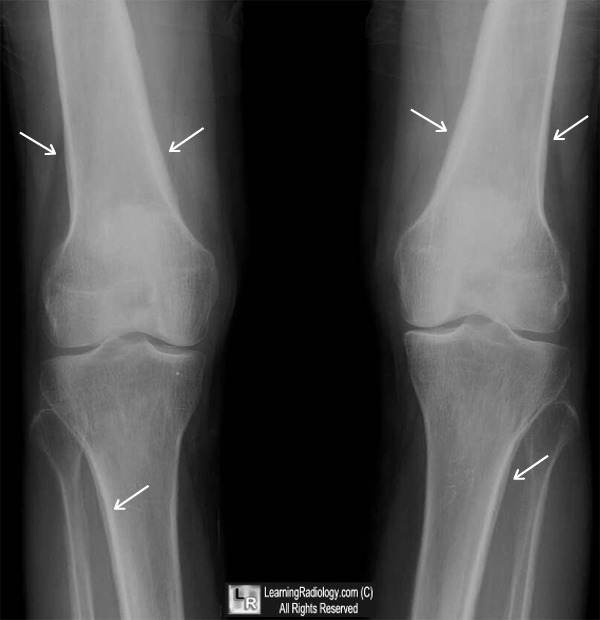
- In diametaphyseal regions
- Periosteal proliferation of new bone
- First smooth, then undulating rough, most conspicuous on concavity of long bones (dorsal and medial aspects)
- Regression of periosteal reaction after thoracotomy
- Soft-tissue swelling ("clubbing") of distal phalanges
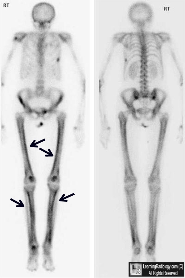
- Bone scan reveals changes early with greater sensitivity and clarity
- Symmetric diffusely increased uptake along cortical margins of diaphysis and metaphysis of tubular bones of the extremities
- Increased periarticular uptake (synovitis)
- Scapular involvement in 2/3
- Mandible ± maxilla abnormal in 40%
|
No comments:
Post a Comment